There is a need for early breast detection to determine early the presence of breast disease. Doctors have long advised women to do BSE (self-examination of breasts) every month. Some tests that are usually carried out to detect cancer or tumors or lumps in the breast are: Ultrasonography (USG), mammography, Automatic Breast Volume Scanner (ABVS) and Magnetic Resonance Imaging (MRI). Each breast radiology examination has advantages and disadvantages. When examining the breast using a 2D ultrasound device, false negatives or errors in the diagnosis of whether the tumor is benign or malignant are often found.
Mammography examinations cannot be carried out on breasts whose tissue is still dense, such as in young women (under 35 years of age), because mammography examinations are carried out by pressing on the breast tissue using a mammography device to obtain an image or pictures with optimal quality in order to increase diagnostic accuracy. Mammography examination can cause discomfort and even pain during the examination. That is why mammography examination is a problem for most women in detecting breast cancer early.
Breast examination using a Magnetic Resonance Imaging (MRI) device even feels more comfortable for women, because the patient only needs to lie on his back on the MRI examination table. The disadvantages of breast examination with MRI are:
It requires quite expensive costs, the examination time is quite long (at least 15-20 minutes) and you feel uncomfortable with the noise of the MRI equipment.
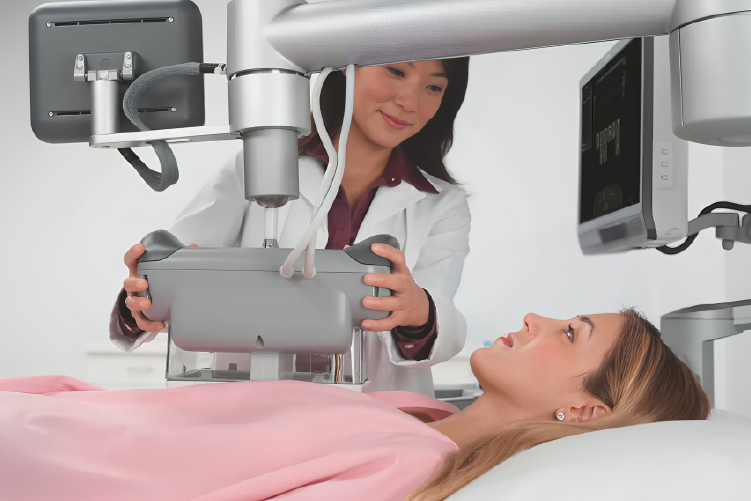
With advances in sophisticated technology, there are now tools for detecting breast tumors, especially in women with dense breast tissue. The examination is relatively painless and uses Ultrasonography (USG) modality so it is safe for everyone, including pregnant women. The tool is known as the Automated Breast Volume Scanner (ABVS).
The Automated Breast Volume Scanner (ABVS) began to be used for breast examinations in Indonesia in 2010. This tool can record three-dimensional images of breast tissue. Data that has been recorded can be analyzed repeatedly. The 3D image can be sliced layer by layer, as thin as 0.5 mm, so that even small tumors can be detected.
Apart from that, ABVS is also equipped with a Doppler system which is useful for distinguishing between benign and malignant tumors through indicators of the level of tissue density and arterial blood flow in the tumor. In general, malignant tumors have sufficient blood flow around the tumor tissue to increase nutrition in the malignant breast tumor tissue.
ABVS has several advantages:
1. Comfort, because the patient is examined in a more comfortable sleeping position with an adjustable level of pressure so that it does not cause pain.
2. Feeling safe, without X-ray radiation, because it uses 5-14 MHz Ultrasonic waves which are safe for the body.
3. Fast, because the breast examination process takes 7-15 minutes.
4. Accurate, because the scan results are three-dimensional slice images from various cross-sections, thus providing adequate data for analysis and diagnosis related to the breast.
5. Details, the thickness of the scanning slices can be adjusted according to the needs related to the patient's condition.
6. Affordable price, because it is not as expensive as an MRI examination.
With these advantages, ABVS examinations can also be carried out on young women and pregnant women. However, it is not recommended for pregnant women to undergo ABVS examinations because it affects the results of the examination.